msk
Tumor Prevalence by Age
| AGE (years) | TUMOR |
| 1 | neuroblastoma |
| 1 - 10 | Ewing's of tubular bones |
| 10 - 30 | osteosarcoma, Ewing's of flat bones |
| 30 - 40 | reticulum cell sarcoma (Primary histiocytic lymphoma), fibrosarcoma |
| | parosteal osteosarcoma, malignant giant cell tumor, lymphoma |
| 40+ | metastatic carcinoma, multiple myeloma, chondrosarcoma |
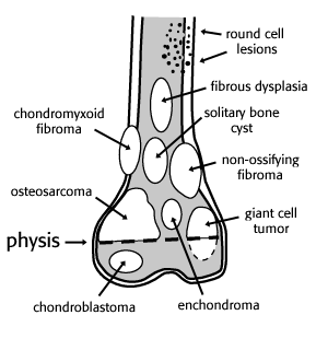
Tumors by Location
Epiphyseal Lesions
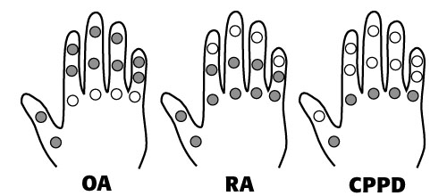
Arthritis Distribution in the Hand
Wrist
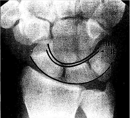
Arcs of Gilula
Carpal Bones
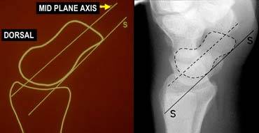
DISI or dorsiflexion instability
DISI is short for dorsal intercalated segmental instability
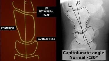
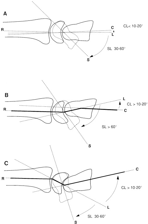
The intercalated segment is the proximal carpal row identified by the lunate. The term 'intercalated segment' refers to it being the part in between the proximal segment of the wrist consisting of the radius and the ulna and the distal segment, represented by the distal carpal row and the metacarpals. In DISI or dorsiflexion instability the lunate is angulated dorsally.
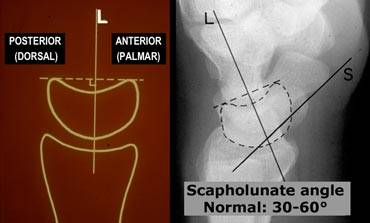
If you think lunate is tilted, measure the scapholunate and capitolunate angle.
VISI or volarflexion instability
Volar intercalated segmental instability or palmar flexion instability is when the lunate is tilted too palmarly. Results in a scapholunate angle of < 30°.
While most DISI is abnormal, in many cases VISI is a normal variant, especially if the wrist is very lax.
SLAC wrist
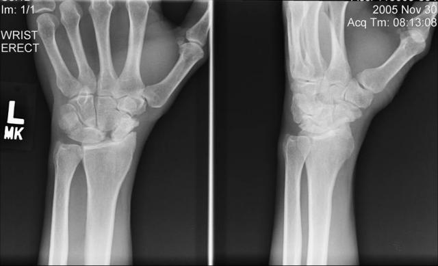
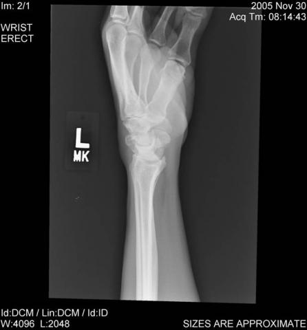
Scapholunate advanced collapse (SLAC) refers to a specific pattern of osteoarthritis and subluxation which results from untreated chronic scapholunate dissociation (scapholunate ligament injury) or from scaphoid non-union (Scaphoid nonunion advanced collapse (SNAC). Other etiologies include Preiser disease (avascular necrosis of the scaphoid), midcarpal instability, intra-articular fractures involving the radioscaphoid or capitate-lunate joints, Kienböck disease, capitolunate degeneration, and inflammatory arthritis, such as seen in the crystalline deposition disorders of gout and calcium pyrophosphate dihydrate deposition disease (CPPD). The radiographic findings of arthropathy in SLAC wrist are nearly identical to those occurring in CPPD.
Wrist radiographs reveal radioscaphoid joint narrowing, sclerosis, osteophytes, cysts, scapholunate dislocation, and carpal collapse. In SLAC secondary to scapholunate dissociation, increased distance between the scaphoid and lunate as well as lunate ulnar translocation will be obvious. A lateral view can show an increase in the scapholunate angle with a dorsiflexion of the lunate (dorsal intercalated segment instability - DISI deformity).
Staging:
Stage 1 A: Narrowing of the radioscaphoid joint first begins at the radial styloid aspect
Stage 1 B: The rest of the radioscaphoid joint is narrowed (the entire scaphoid fossa is involved).
Stage 2: The capitolunate joint is additionally narrowed and sclerotic. This results in a radial or dorsal radial position of the capitate (midcarpal SLAC).
Scoliosis
DDx
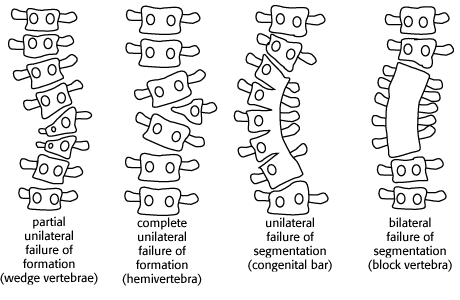
congenital (secondary to vertebral anomalies, which may have associated tethered cord or diastematomyelia)
neuromuscular
diseases of collagen synthesis
neurofibromatosis
tumors (especially osteoid osteoma)
radiation injury
Vertebral Anomalies Causing Scoliosis
DDx of Soft Tissue Calcifications
| Cause | Typical Appearance | Prevalence |
| Dystrophic | small to large amorphous Ca++ in the damaged tissue – may progress to ossification (formation of cortex and medullary space are then seen) | 95 - 98 % |
| CPPD | chondrocalcinosis; occasionally associated with calcifications in the soft tissues of the spine | 1 - 2 % |
| Metastatic calcification | finely speckled Ca++ throughout soft tissues | 1 - 2 % |
| Tumoral calcinosis | big globs of Ca++, usually near a joint | << 1 % |
| Metastatic osteosarcoma | amorphous, fluffy, confluent collection of Ca++ | <<< 1 % |
| Primary soft tissue osteosarcoma | amorphous, fluffy, confluent collection of Ca++ | <<<< 1 % |
DDx Periostial Reaction
DDx Acroosteolysis
DDx Vertebra Plana (FETISH)
Fracture
EG
Tumor (leukemia,NB)
Infection
Steroids
Hemangioma
Causes of AVN
Stages of AVN
| Stage | Findings |
| 0 | asymptomatic, normal radiographs |
| I | normal radiographs (abnormal MRI) |
| II | radiolucency and sclerosis |
| III | crescent sign, normal contour |
| IV | subchondral collapse, flattening |
| V | degenerative joint disease |
Trevor-Fairbanks Disease
Intraarticular osteochondromas arising from the epiphysis
May occur in single or multiple epiphyses and generally is found on only one side of the body
Knee and the ankle are the most common sites
Histologically identical to an exostosis
Cystic Soft Tissue Tumors
Ganglion Cyst
Synovial Cyst
Cystic Schwannoma
Synovial Sarcoma
Mucoid Liposarcoma
Myxomatous Tumors
Paget's Degenerates Into
MFH
Osteosarcoma
Giant Cell
McCune-Albright
Mazabraud Syndrome
Common Locations for Insufficiency Fractures
Sacrum
Femoral Neck
Pubic Rami
Spine
Soft Tissue Calcifications in the Hand
Arthritis w/ SI Involvement
Ivory Vertebrae
Lymphoma
Paget's
Sclerotic Met
AC Separation
Grade I
Grade II
Grade III
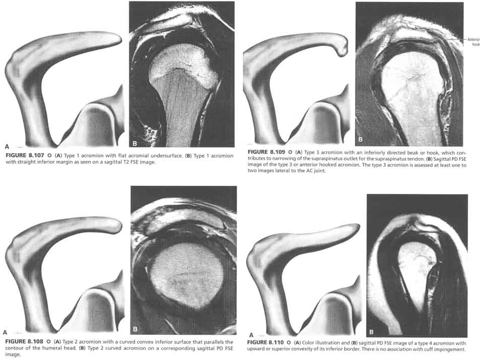
Acromion Types
DDx Deep Muscle Edema
Pes Anserine Bursitis
pes anserine bursa is proximal and medial to the primary attachment of the medial hamstrings on the proximal tibia
medial hamstrings that form the roof of the pes anserine bursa are the sartorius, gracilis, and semitendinosus (SGT)
Soft Tissue Tumors of Hand
Charcot Joint
5 “D’s”
Density → increased or normal
Distension → effusion
Debris → bone fragments
Disorganization
Dislocation
40% of Charcot is the primary atrophic type in which you don’t have the 4 D’s, just resorption of bone
Etiology → secondary to loss of pain perception and altered sympathetic control of blood flow
diabetes
alcoholism
nerve/spinal cord injury
polio
neurosyphilis
syrinx
myelomeningocele
steroids
scleroderma
Charcot Spine
usually secondary to tabes dorsalis or syrinx
increased density, deformity, fragmentation, and dislocation
destruction usually involves the intervertebral disc as well as the vertebral body
Sinus Tarsi Syndrome
Calcaneal Pitch
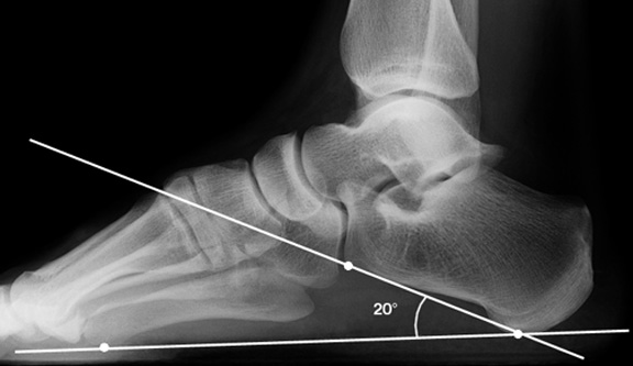
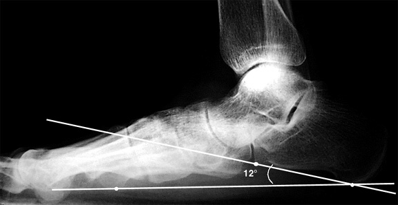
A line is drawn from the plantar-most surface of the calcaneus to the inferior border of the distal articular surface. The angle made between this line and the transverse plane (or the line from the plantar surface of the calcaneus to the inferior surface of the 5th metatarsal head) is the calcaneal pitch. A decreased calcaneal pitch is consistent with pes planus.
18 to 20° is generally considered normal, although measurements ranging from 17 to 32° have been reported to be normal.
Hallux Valgus
Sesamoids
Moderate subluxation
Severe subluxation
Lisfranc Ligaments
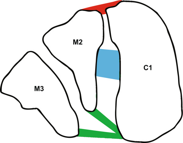
C1 = Medial cuneiform
M2 = 2nd metatarsal base
M3 = 3rd metatarsal base
Red = dorsal Lisfranc ligament
Blue = interosseous Lisfranc ligament
Green = plantar Lisfranc ligament
Patella Alta
Insall-Salvati Ratio
ratio of the patella tendon length (TL) to the length of the patella (PL). This can be measured on a lateral knee xray or sagittal MRI. Ideally the knee is 30 degrees flexed.
The traditional number used to TL:PL is < 1.2 (between 0.8 and 1.2), although more recent literature 2 suggests that the true range of normal is more forgiving - up to 0.74 to 1.50.
Patellar length (PL) is the greatest pole - pole length
Patellar tendon length (TL) is defined as the length of the post surface of the tendon from the lower pole of patella to its insertion on the tibia.
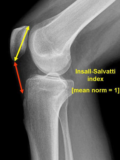
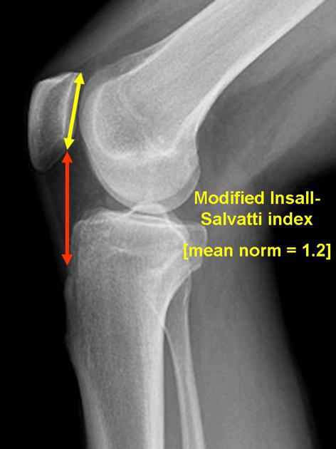
Modified Insall-Salvati Ratio
Mnemonics
Dense Bones (3M's PROOF)
Mets
Myelofibrosis
Mastocytosis
Sickle Cell
Pyknodysostosis
Renal Osteodystrophy
Osteopetrosis
Others
Fluorosis
DDx Multiple Lucent Bone Lesions (FEMHI)
MSK Interventional
Shoulder Arthrogram
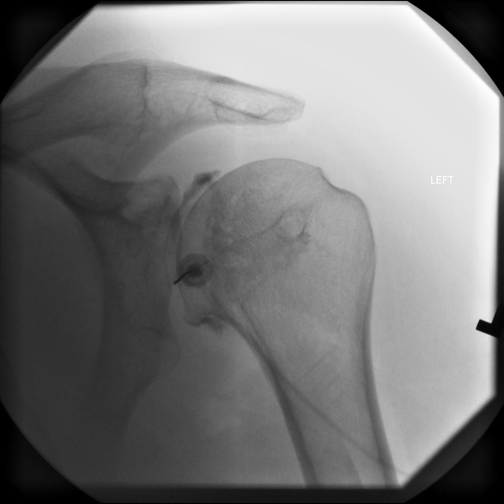
Use 22 G spinal needle
MR Arthrogram:
Inject 12 cc of a solution of 5 cc normal saline, 5 cc Omnipaque 300, 10 cc 1% lidocaine, and 0.1 cc gadolinium
CT Arthrogram:
msk.txt · Last modified: 2024/07/16 15:47 by 127.0.0.1

